
Jennifer couldn’t explain what was wrong.
Her knee didn’t hurt, exactly. It just felt… off. Unstable. Like her leg might give out at any moment.
She’d been to three doctors. Had two MRIs. Both showed minor wear and tear—nothing that explained why a former college athlete suddenly couldn’t trust her own leg to walk down stairs.
“They keep telling me my knee is fine,” she said during her first visit to our Ashland chiropractic office. “But something is clearly not working.”
She was right. Something wasn’t working.
But it wasn’t her knee.
It was the information loop between her knee and her brain—a four-step circuit that controls everything from how you walk to whether you feel pain, how strong your muscles contract to how coordinated your movements are.
And when that loop breaks down, even a structurally “fine” joint can feel completely unreliable.
“Let me show you something,” I said, pulling out a diagram. “Your knee isn’t the problem. Your nervous system has stopped listening to it.”
The Hidden Control System Running Your Body
Most people think of their nervous system as a simple on/off switch: brain sends signal → muscle contracts → movement happens.
But that’s not how it works.
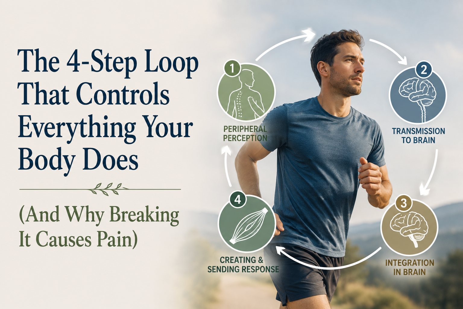
Your nervous system operates as a continuous feedback loop—a four-step cycle that runs thousands of times per second, controlling every movement, every sensation, and every protective response your body generates.
Here’s the loop:
Step 1: Peripheral Perception
Specialized receptors throughout your body detect what’s happening in your internal and external environment.
Step 2: Transmission to Brain
These signals travel up sensory nerves through your spinal cord to your brain.
Step 3: Integration in Brain
Your brain processes, integrates, and interprets these signals, then decides how to respond.
Step 4: Creating & Sending Response
Your brain sends output signals back down through your spinal cord to your muscles, organs, or wherever action is needed.
Then the loop starts over. Immediately.
This happens so fast and so automatically that you’re not consciously aware of it. But this loop is literally running your life.
And when any step in this loop breaks down?
That’s when pain, weakness, instability, and dysfunction show up—even when your structure looks “fine” on imaging.
Step 1: Peripheral Perception—Your Body’s Sensor Network
The first step of the loop is where your body gathers information about what’s happening right now.
You have millions of specialized sensors embedded throughout your tissues, constantly monitoring your internal and external environment:
Mechanoreceptors in your joint capsules detect:
- Position and movement (proprioception)
- Pressure and compression
- Stretch and tension
- Vibration
Muscle Spindles embedded within your muscle fibers track:
- How long or short the muscle is
- How fast the muscle length is changing
- Whether the muscle is being stretched
Golgi Tendon Organs (GTOs) located in your tendons measure:
- How much force the muscle is producing
- How much tension is on the tendon
- Whether the load is approaching dangerous levels
Thermoreceptors sense:
- Temperature changes in your tissues
- Heat or cold in your environment
Chemoreceptors monitor:
- Chemical changes in your blood and tissues
- pH levels
- Oxygen and carbon dioxide concentrations
- Inflammatory markers
Baroreceptors detect:
- Blood pressure changes
- Vascular stretch
Your Five Special Senses:
- Vision (light and spatial information)
- Hearing (sound and balance via the vestibular system)
- Smell (chemical detection in air)
- Taste (chemical detection in food)
- Touch (pressure, texture, pain receptors in skin)
All together, these sensors are feeding your nervous system approximately 11 million bits of information per second.
That’s 11 million data points about where you are, what you’re doing, and whether you’re safe.
And this is just Step 1 of the loop.
Step 2: Transmission to Brain—The Information Highway
Once your sensors detect something, that information needs to get to your brain.
This is where your sensory nerves come in.
These nerves act like fiber optic cables, carrying electrical signals from your periphery up through your spinal cord to your brainstem and ultimately to your brain.
But not all information travels at the same speed.
Fast-conducting fibers (A-beta fibers) carry information about:
- Light touch
- Vibration
- Proprioception (joint position)
These signals travel at speeds up to 120 meters per second—almost as fast as a Major League fastball.
Slower-conducting fibers (A-delta and C fibers) carry information about:
- Pain
- Temperature
- Deep pressure
These travel at 1-30 meters per second—still fast, but significantly slower than proprioceptive signals.
Here’s why this matters:
The quality and clarity of the signal going up determines the quality of the response coming back down.
If your joint mechanoreceptors aren’t firing properly (because a joint is stiff or immobile), your brain receives blurry, incomplete information.
If your muscle spindles are hypersensitive (because of chronic guarding), your brain gets distorted data about muscle length and tension.
If your signals are delayed or degraded (because of nerve compression or poor tissue health), your brain is working with outdated or inaccurate information.
And here’s the critical part:
Your brain can only make decisions based on the information it receives. Garbage in, garbage out.
This is what was happening with Jennifer’s knee.
Step 3: Integration in Brain—Where Decisions Get Made
Once the sensory information arrives at your brain, something extraordinary happens:
Your brain doesn’t just passively receive this information. It actively predicts what the information means and what to do about it.
Remember from our earlier nervous system series: your brain operates as a prediction machine (thanks to Lisa Feldman Barrett’s work).
It’s constantly running simulations based on past experience, asking:
- “What does this pattern of input mean?”
- “Is this dangerous?”
- “What should I do about it?”
This integration happens across multiple brain regions simultaneously:
The Thalamus acts as a relay station, routing sensory information to appropriate processing centers.
The Somatosensory Cortex creates and maintains a map of your body—telling you where sensations are coming from and what they mean.
The Interoceptive Network (including the insula and anterior cingulate cortex) generates your felt sense of your body’s internal state—the sensations that create your experience of comfort, discomfort, pain, or ease.
The Motor Cortex plans and initiates voluntary movements.
The Cerebellum coordinates complex movements and maintains balance and timing.
The Limbic System evaluates emotional significance and threat level.
The Prefrontal Cortex makes executive decisions about how to respond.
All of this processing happens in milliseconds.
Your brain integrates 11 million bits of incoming sensory data, compares it against past experience and predictions, evaluates threat level, and generates a coordinated response.
And then comes Step 4.
Step 4: Creating & Sending Response—The Output
Based on all the information processed in Step 3, your brain creates an output signal and sends it back down through your spinal cord to your muscles, organs, or glands.
This output could be:
Motor commands to muscles:
- “Contract the quadriceps to stabilize the knee”
- “Relax the hamstrings to allow the leg to straighten”
- “Fire the hip abductors to keep the pelvis level”
Autonomic responses to organs:
- Increase heart rate
- Redirect blood flow to muscles
- Trigger stress hormone release
- Initiate digestive processes
Pain signals:
- Generate discomfort to warn of potential danger
- Create protective muscle guarding
- Limit range of motion
Coordination patterns:
- Sequence muscle activation for smooth movement
- Adjust balance and posture
- Modulate force output
These output signals travel down motor nerves at speeds up to 120 meters per second, telling your body exactly what to do.
And here’s what most people don’t realize:
The quality of your output is entirely dependent on the quality of your input.
You can have the strongest muscles in the world, but if your brain receives poor proprioceptive input from stiff joints, it will limit motor output to protect you.
You can have perfect structural anatomy, but if your muscle spindles are sending distorted signals, your brain will generate dysfunctional movement patterns.
You can do all the “right” exercises, but if your nervous system loop is broken at Step 1 or Step 2, your brain will never generate the outputs you’re trying to achieve.
Where Jennifer’s Loop Broke Down
When we tested Jennifer’s knee, here’s what we found:
Step 1 was broken. Several segments in her lumbar spine were stiff and hypomobile. This meant the mechanoreceptors in those joint capsules weren’t firing properly, creating poor proprioceptive input.
Additionally, her knee joint itself had reduced mobility from months of guarding. More silent mechanoreceptors.
Step 2 was degraded. The poor-quality signals from her spine and knee were traveling up to her brain, but they were blurry, incomplete, and delayed.
Step 3 became distorted. Her somatosensory cortex—the brain’s map of her body—had become “smudged” around her knee and lower back. Her brain literally didn’t have a clear picture of where her leg was in space or how it was moving.
Step 4 was protective. Based on unclear input and a distorted body map, her brain’s prediction was: “This knee is unstable. Movement is dangerous.”
So it generated protective outputs:
- Excessive muscle guarding in her quadriceps and hamstrings
- Limited motor unit recruitment
- Reduced force production
- The subjective feeling of instability
Her knee wasn’t structurally unstable.
Her nervous system just thought it was.
And that perception became her reality.
How We Fixed the Loop
Fixing Jennifer’s knee meant fixing the loop—starting at Step 1.
Restore Input Quality (Step 1)
We used specific chiropractic adjustments to restore motion to the stiff segments in her lumbar spine and improve mobility in her knee joint itself.
Every time a joint moves properly, it activates mechanoreceptors and sends a flood of high-quality proprioceptive signals to the brain.
We also used targeted manual therapy on her muscle spindles and GTOs—recalibrating their sensitivity so they stopped sending distorted signals about muscle length and tension.
Improve Signal Transmission (Step 2)
We incorporated nerve gliding exercises for her femoral nerve and tibial nerve—gentle movements that improve nerve mobility and signal conduction.
We also used the Neubie with Direct Current to flood her nervous system with clear, directional proprioceptive input that her brain could actually understand and use.
Remap the Brain (Step 3)
We used slow, controlled movements with eyes closed to help Jennifer’s brain redraw its somatosensory map of her knee.
We practiced:
- Single-leg balance with focus on precise knee position
- Controlled squatting with attention to where her knee tracked
- Step-downs with emphasis on smooth, coordinated movement
Each successful repetition updated her brain’s prediction: This is safe. You can trust this knee.
Allow Better Outputs (Step 4)
As her input quality improved and her brain’s map became clearer, her motor outputs naturally improved.
Her quadriceps and hamstrings learned to coordinate instead of fighting each other.
Her nervous system allowed fuller motor unit recruitment.
Her subjective sense of instability disappeared.
Not because we “strengthened” anything.
Because we fixed the loop.
The Loop in Action: Why “Normal” MRIs Can Still Hurt
This is why imaging so often misses the real problem.
An MRI shows hardware—bones, cartilage, ligaments, discs.
But it doesn’t show:
- Whether your mechanoreceptors are firing
- How clear your proprioceptive signals are
- What your somatosensory cortex looks like
- How your brain is predicting threat
You can have a “perfect” MRI and still have a broken loop.
And when the loop is broken, you’ll have:
- Pain that doesn’t match your imaging
- Weakness that doesn’t match your muscle size
- Instability that doesn’t match your structural integrity
- Dysfunction that no one can explain
Because the problem isn’t in your tissues.
It’s in the information flow between your tissues and your brain.
The Three Most Common Places the Loop Breaks
In our Ashland chiropractic practice, we see the loop break down in three predictable patterns:
1. The Input Problem (Steps 1-2)
Stiff joints → Silent mechanoreceptors → Blurry proprioceptive signals → Poor brain mapping → Protective motor output
Fix: Restore joint mobility, recalibrate muscle spindles, improve nerve conduction
2. The Integration Problem (Step 3)
Chronic pain → Distorted somatosensory maps → Inaccurate body representation → Threat-based predictions → Protective motor output
Fix: Remap the brain through movement education, reduce threat perception, improve prediction accuracy
3. The Output Problem (Step 4)
Even with good input, the brain generates protective outputs due to:
- Learned fear-avoidance
- Previous injury patterns
- Sympathetic nervous system dominance
Fix: Graded exposure to safe movement, parasympathetic activation, reframe pain understanding
Most chronic pain and movement dysfunction involves all three.
That’s why a comprehensive approach—one that addresses the entire loop—is so much more effective than isolated treatments that only target one step.
Jennifer’s Breakthrough
After eight weeks of working on her nervous system loop, Jennifer was back to running.
Not jogging. Running. Five miles at a time, pain-free, with complete confidence in her knee.
Her knee structure hadn’t changed. The “wear and tear” on her MRI was still there.
But her nervous system loop was working again.
Clear input → Accurate transmission → Precise integration → Coordinated output.
The loop was whole. And when the loop works, the body works.
Your Body Is Smarter Than Your Imaging
If you’ve been told your imaging looks “fine” but you still have pain, weakness, or instability…
If you’ve done all the “right” exercises but nothing seems to stick…
If doctors keep saying “we can’t find anything wrong”…
Consider this: they’re looking at your hardware.
But your problem might be in your software—in the four-step loop that controls everything your body does.
And when you fix that loop?
Everything else can finally fall into place.
That’s what we do every day at our Ashland chiropractic office—identifying where your nervous system loop has broken down and systematically restoring each step until information flows clearly again.
Not forcing your body to change.
But giving your nervous system the clarity it needs to allow change.
Because sometimes the answer isn’t in your joints or your muscles or your imaging.
It’s in the loop.
References
- Proske U, Gandevia SC. The proprioceptive senses: their roles in signaling body shape, body position and movement, and muscle force. Physiol Rev. 2012;92(4):1651-1697.
- Tuthill JC, Azim E. Proprioception. Curr Biol. 2018;28(5):R194-R203.
- Brumagne S, Cordo P, Lysens R, et al. Role of paraspinal muscle spindles in lumbosacral position sense. Spine. 2000;25(8):989–94.
- Flor H, Braun C, et al. Extensive reorganization of primary somatosensory cortex in chronic back pain patients. Neurosci Lett. 1997;224(1):5–8.
- Moseley GL. I can’t find it! Distorted body image and tactile dysfunction in patients with chronic back pain. Pain. 2008;140(1):239-43.
- Hodges PW, Tucker K. Moving differently in pain: a new theory to explain the adaptation to pain. Pain. 2011;152(3 Suppl):S90-8.
- Barrett LF. How Emotions Are Made: The Secret Life of the Brain. Houghton Mifflin Harcourt; 2017.
- Purves D, Augustine GJ, Fitzpatrick D, et al. Neuroscience. 6th edition. Sunderland (MA): Sinauer Associates; 2017.